Upper Inner Thigh Anatomy - How To Prevent Groin Injuries : Medial compartment, also known as adductor compartment;. People who play soccer have these specific muscles of the leg very well defined, so they're like a walking anatomy atlas for thigh muscles. Damage, disruption or injury to any of its components can result in dully, achy thigh pain. The fat underneath that loose leg skin actually provides structure to the overlying skin. The thigh bears much of the load of the body's weight when a person is upright. The thigh muscles don't just move your legs.
It transmits the great saphenous vein, and other, smaller vessels, and is termed the fossa ovalis. Meanwhile, the vastus lateralis is on the side of the thigh, while the vastus intermedius is hidden below the rectus femoris(5). The thigh bears much of the load of the body's weight when a person is upright. Pain in the groin and. The femur is the longest and strongest bone in the body.
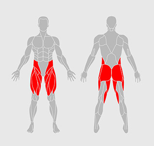
Inner Thighs Workout from darebee.com On the anterior side, the most prominent of the muscles are the sartorius muscle and the four muscles that make up quadriceps muscle group (the quads.) The femur is the longest and strongest bone in the body. The thigh bears much of the load of the body's weight when a person is upright. The gracilis muscle runs down the surface of your inner thigh from between your groin and into the inner knee. The external iliac artery supplies blood to the femoral artery. Anterior muscles extend your legs and flex your thighs. Started in 1995, this collection now contains 6907 interlinked topic pages divided into a tree of 31 specialty books and 737 chapters. Medial compartment, also known as adductor compartment;
Started in 1995, this collection now contains 6907 interlinked topic pages divided into a tree of 31 specialty books and 737 chapters.
All the medial thigh muscles are innervated by the obturator nerve, which arises from the lumbar plexus. Cramps in the inner thigh, or groin, are also common. The following diagram illustrates the actions of the terms adduction, abduction, flexion and extension at the different joints. People who play soccer have these specific muscles of the leg very well defined, so they're like a walking anatomy atlas for thigh muscles. Anterior muscles extend your legs and flex your thighs. The upper third of the artery is contained in the femoral triangle, which is also known as the scarpa's triangle, and its middle third is contained in the hunter's canal, or the adductors canal, as it is commonly called. In many instances, i see surgeons took too much fat out of the upper thigh without addressing the skin issue. Here, it lies midway between the anterior superior iliac spine and the symphysis pubis. The thigh muscles don't just move your legs. The thigh has three sets of strong muscles: Muscle anatomy abdominal region 12 photos of the muscle anatomy abdominal region muscle anatomy abdominal region, human muscles, muscle anatomy abdominal region. The thigh contains one major bone and many muscles, nerves, and arteries; The femoral nerve supplies the skin around the inner leg and the upper thigh area, the saphenous nerve supplies the medial aspect of the lower leg—a small area on of the foot and the ankle.
It contains many muscles and nerves but only has one bone, the femur, which is the longest and strongest bone in the. The external iliac artery supplies blood to the femoral artery. The femur is the longest and strongest bone in the body. The sciatic nerve also referred to as the largest nerve in the human body, courses from the lower back down the lower extremity. The femoral artery enters the thigh from behind the inguinal ligament as the continuation of the external iliac artery.
Anatomy Of Liposuction Of Theabdomen Hips Thighs Aesthetic Surgery from www.78stepshealth.us They have a lot to do with how your hips move. At the top of the thigh, it passes through the saphenous opening of the fascia lata and enters the deeper tissues of the upper thigh before merging into the femoral vein. It is present in the lower abdomen but goes up to the upper inner thigh. These muscles sit close to the groin, which refers to the region of the hip between the stomach and thigh. Medial muscles adduct and rotate your thigh, and posterior flex your leg and extend your thigh. It can also make your clothing fit uncomfortably, and hinder the appearance of long, lean legs. Anterior compartment, also known as the extensor compartment; The muscles of the thigh and gluteal region are a group of complex muscles that help move and stabilize the lower limb.
The thigh contains one major bone and many muscles, nerves, and arteries;
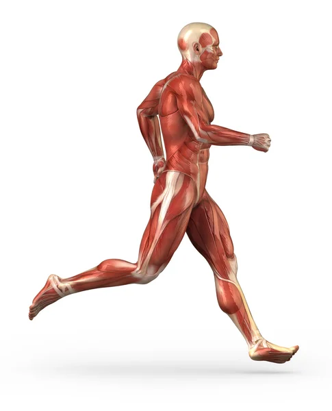
What causes dull, achy thigh pain? Here, it lies midway between the anterior superior iliac spine and the symphysis pubis. The upper third of the artery is contained in the femoral triangle, which is also known as the scarpa's triangle, and its middle third is contained in the hunter's canal, or the adductors canal, as it is commonly called. Is there an easy way to learn their a. Started in 1995, this collection now contains 6907 interlinked topic pages divided into a tree of 31 specialty books and 737 chapters. The muscles in the medial compartment of the thigh are collectively known as the hip adductors. In clinical anatomy the thigh muscles are divided into three groups: It is present in the lower abdomen but goes up to the upper inner thigh. Like the forearm, the upper leg, or thigh, has a dense arrangement of many muscles. This small fat compartment located in the upper thigh is responsible for excessive friction when you walk, run, or engage in other activities. The quadriceps and hamstrings work together to straighten (extend) and bend (flex) the leg. The femoral artery enters the thigh from behind the inguinal ligament as the continuation of the external iliac artery. The muscles of the thigh and gluteal region are a group of complex muscles that help move and stabilize the lower limb.
Medial compartment, also known as adductor compartment; The bone of the thigh is called the femur. It is present in the lower abdomen but goes up to the upper inner thigh. There, the nerve divides into its anterior and posterior branches. What causes dull, achy thigh pain?
ሠUpper Inner Thigh Massage Stock Photos Royalty Free Adductor Images Download On Depositphotos from st.depositphotos.com The upper inner thigh tends to have very thin, loose skin for many patients, both men and women. Is there an easy way to learn their a. Anterior muscles extend your legs and flex your thighs. The adductor muscles pull the legs together. This small fat compartment located in the upper thigh is responsible for excessive friction when you walk, run, or engage in other activities. Related posts of muscle anatomy of upper thigh muscle anatomy abdominal region. The thigh contains one major bone and many muscles, nerves, and arteries; Meanwhile, the vastus lateralis is on the side of the thigh, while the vastus intermedius is hidden below the rectus femoris(5).
The gracilis muscle runs down the surface of your inner thigh from between your groin and into the inner knee.
The hamstring muscles in the back of the thigh, the quadriceps muscles in the front, and the adductor muscles on the inside. The thigh bears much of the load of the body's weight when a person is upright. There are five muscles in this group; The gracilis muscle runs down the surface of your inner thigh from between your groin and into the inner knee. The upper third of the artery is contained in the femoral triangle, which is also known as the scarpa's triangle, and its middle third is contained in the hunter's canal, or the adductors canal, as it is commonly called. Pain in the groin and. The femoral nerve supplies the skin around the inner leg and the upper thigh area, the saphenous nerve supplies the medial aspect of the lower leg—a small area on of the foot and the ankle. The adductor muscles pull the legs together. The thigh is the region between the hip and knee joints. Anterior muscles extend your legs and flex your thighs. All the medial thigh muscles are innervated by the obturator nerve, which arises from the lumbar plexus. The thigh has three sets of strong muscles: These work with the quadriceps, hamstrings, buttocks and deep hip rotators to rotate your leg.
Related posts of muscle anatomy of upper thigh muscle anatomy abdominal region upper thigh anatomy. Continuing through the thigh, the great saphenous vein turns anteriorly while merging with several more superficial veins.

0 Comments:
Post a Comment